Home

méthodologie révolution après ça mitochondria light microscope Palais des enfants Harmonie contre

Why can't we see cell organelles such as mitochondria, ribosomes, plastics,  etc under a compound microscope, although it is stained darker than  cytoplasm? - Quora
Why can't we see cell organelles such as mitochondria, ribosomes, plastics, etc under a compound microscope, although it is stained darker than cytoplasm? - Quora

Imaging captures rogue molecule that can trigger autoimmunity | Centre for  Dynamic Imaging
Imaging captures rogue molecule that can trigger autoimmunity | Centre for Dynamic Imaging

Plant Mitochondria Viewed Under Microscope Stock Photo 1241522146 |  Shutterstock
Plant Mitochondria Viewed Under Microscope Stock Photo 1241522146 | Shutterstock

Mitochondria: A worthwhile object for ultrastructural qualitative  characterization and quantification of cells at physiological and  pathophysiological states using conventional transmission electron  microscopy - ScienceDirect
Mitochondria: A worthwhile object for ultrastructural qualitative characterization and quantification of cells at physiological and pathophysiological states using conventional transmission electron microscopy - ScienceDirect

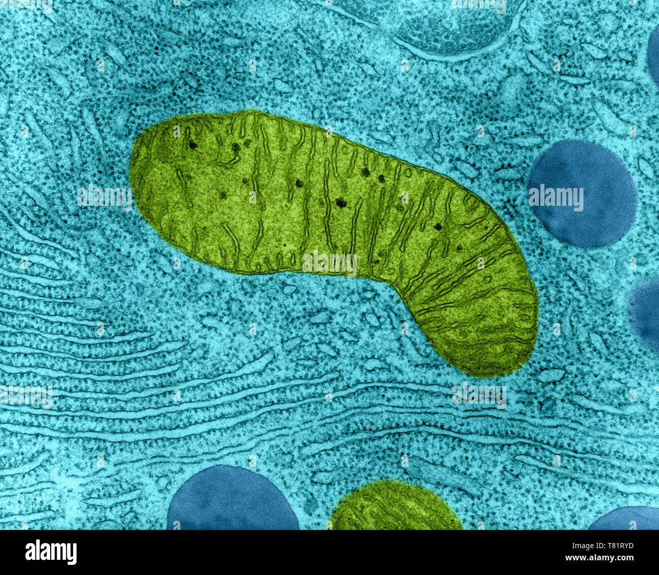
Tem mitochondria hi-res stock photography and images - Alamy

Live mitochondria seen in unprecedented detail: photobleaching in STED  microscopy overcome
Live mitochondria seen in unprecedented detail: photobleaching in STED microscopy overcome

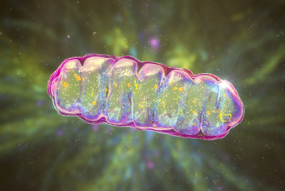
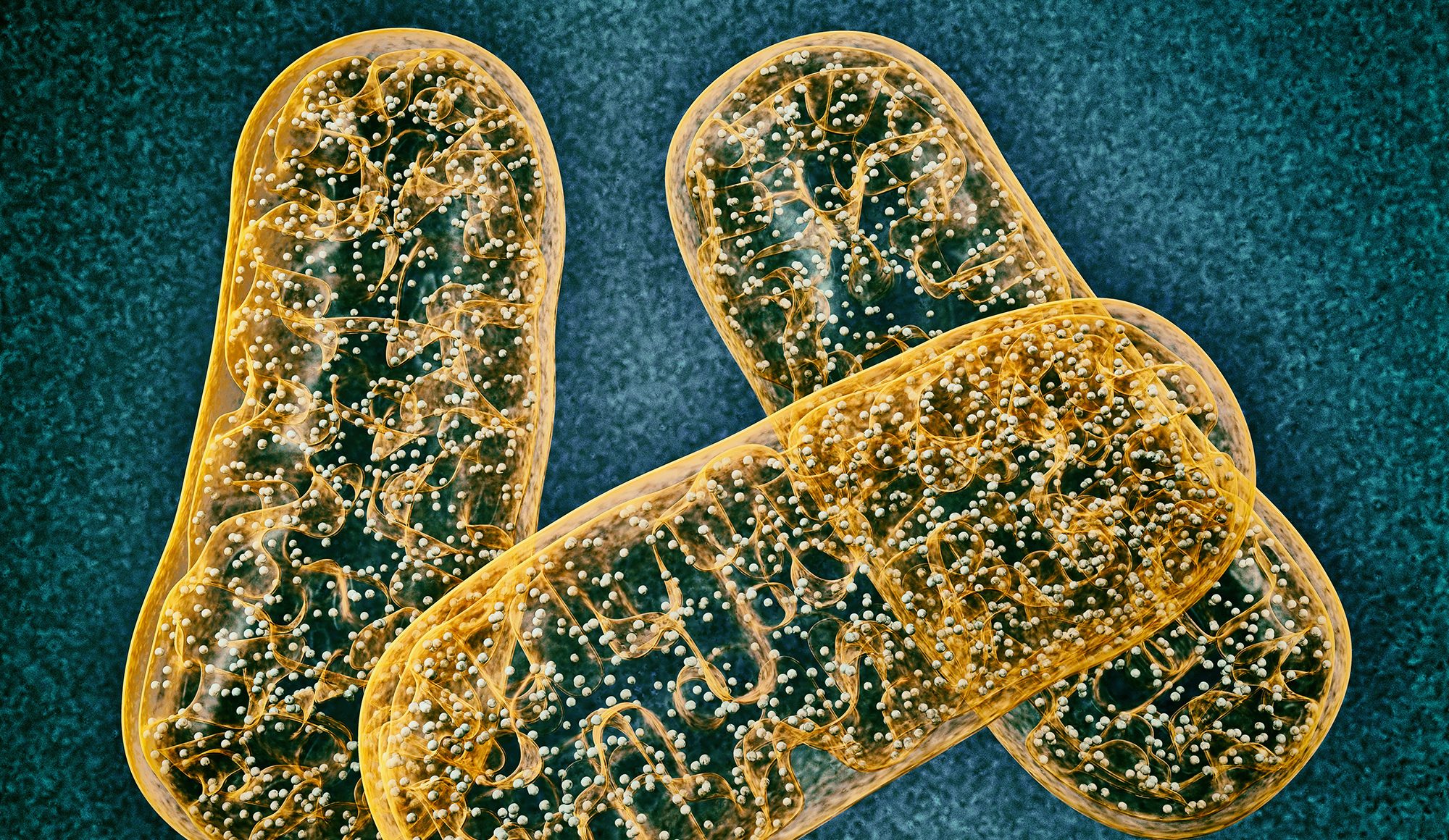
Mitochondria Under Microscope On Dark Blue Backgound In Futuristic Glowing  Low Polygonal Style Stock Illustration - Download Image Now - iStock
Mitochondria Under Microscope On Dark Blue Backgound In Futuristic Glowing Low Polygonal Style Stock Illustration - Download Image Now - iStock

Nagoya University Research Achievements

Mitochondria under the microscope — Science Learning Hub

Mitochondria appearance under electron microscope (EM × 6000); A :... |  Download Scientific Diagram
Mitochondria appearance under electron microscope (EM × 6000); A :... | Download Scientific Diagram

Study sheds light on a mitochondrial disease - News - University of  Liverpool
Study sheds light on a mitochondrial disease - News - University of Liverpool

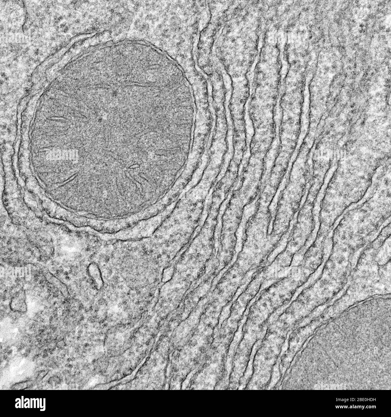
Mitochondria micrograph hi-res stock photography and images - Alamy

Ultrastructural examination of ER−mitochondria tethering in HEK 293T... |  Download Scientific Diagram
Ultrastructural examination of ER−mitochondria tethering in HEK 293T... | Download Scientific Diagram
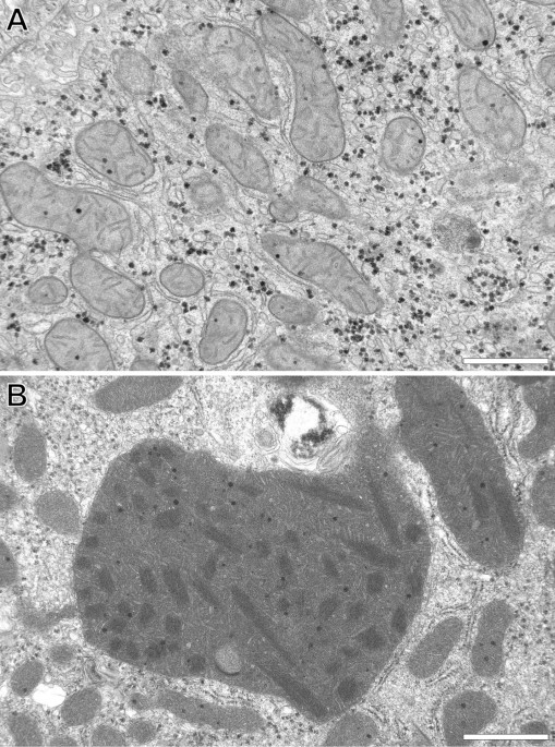
Insight into mitochondrial structure and function from electron tomography  - ScienceDirect
Insight into mitochondrial structure and function from electron tomography - ScienceDirect

Three-dimensional ultrastructure of giant mitochondria in human  non-alcoholic fatty liver disease | Scientific Reports
Three-dimensional ultrastructure of giant mitochondria in human non-alcoholic fatty liver disease | Scientific Reports

Light microscopy Images of clusters of isolated mitochondria on mica at...  | Download Scientific Diagram
Light microscopy Images of clusters of isolated mitochondria on mica at... | Download Scientific Diagram

Light and electron microscopy showing ultrastructural changes in the... |  Download Scientific Diagram
Light and electron microscopy showing ultrastructural changes in the... | Download Scientific Diagram

The morphology of mitochondria. (a) Thin-section electron micrograph of...  | Download Scientific Diagram
The morphology of mitochondria. (a) Thin-section electron micrograph of... | Download Scientific Diagram

Mechanisms in Aging Process Illuminated by Light-Powered Mitochondria

Mitochondria, TEM - Stock Image - C057/6555 - Science Photo Library

Correlated three-dimensional light and electron microscopy reveals  transformation of mitochondria during apoptosis | Nature Cell Biology
Correlated three-dimensional light and electron microscopy reveals transformation of mitochondria during apoptosis | Nature Cell Biology

Mitochondria Under Electron Microscope - YouTube

Mitochondrial swelling detection with a bright-field microscope.... |  Download Scientific Diagram
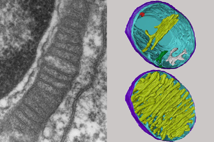
Mitochondrial swelling detection with a bright-field microscope.... | Download Scientific Diagram

Worms Live Longer with Mitochondria Powered by Light: Preprint | The  Scientist Magazine®
Worms Live Longer with Mitochondria Powered by Light: Preprint | The Scientist Magazine®

Are mitochondria the natural cellular light sources?